How do you pack cord?
Packing cord is a probably a dying art, but if margins are subgingival and they need to be captured , the gingiva needs retraction. To properly pack cord, it's best to get any bleeding to stop before attempting to pack cord. If the sulcus is too moist it is much harder getting the cord to stay in the sulcus ( it tends to float out when it is overly wet).
Placing a finish line subgingivally often causes bleeding, since the gingiva can be abraded. I personally like creating one mm long bevels sub gingivally especially on the proximal portions of my preparations and this definitely tends to cause bleeding. If the finish line is placed only .5 mm subgingivally, impressioning and cord packing is usually easier, but if there is any appreciable gingival recession the margin of my restoration becomes visible ( not a good thing with ceramometal restorations). To stop any bleeding I use Ultradent's Viscostat ( a ferric sulfate preparation that is gentle for the gingiva). It is applied with an applicator supplied by the company and rubbed gently and repeatedly into the sulcus. The gentle rubbing motion is followed by rinsing with water ( without the airspray) is used to remove the excess. I tend to use a two by two cotton as a barrier to keep this material from getting on the tongue (It tastes really bad!). After an application, I set a timer and wait 4 minutes. I then gently wash the sulcus again. If there is still some bleeding I will reapply the viscostat and rewash. Usually this works to stop any slight renewed bleeding.
Once the gingiva is relatively dry, I am ready to pack cord. I Start by tacking down the first piece . Usually this is easier when placed at a line angle (one of the corners) first. I press down on the cord with the cord packer, perpendicular to the cord, and hold with a gentle but firm pressure for a couple of seconds. If the cord is not overly wet and the "sulcus" is intact, the cord will probably stay put. If the cord is much thinner than the width of the sulcus it may float out any way because the gingiva adjacent to it will not hold on the cord. It is easiest to start the cord in a part of the sulcus that is snug enough to hold onto the cord. Once the first section is "tacked down" I slowly work my way around my preparation with a gentle but firm pressure. Each time I push a new section down, I wait a second or two before removing my cord packer. The force applied must be either vertical or pointed slightly towards the previously packed piece of cord. This insures that the cord packer will not pull on the part that was previously placed.
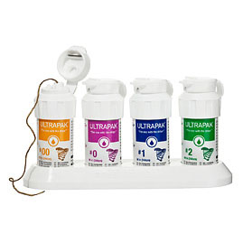
I pack two layers of cord, the first a thinner cord (ultradent 0) and the second thicker (ultradent 1). After I place both my cords I place a wet two by two over the prep, ask my patient to bite down and set my timer for another 4 minutes. Waiting allows my patients naturally clotting mechanism the time it needs to work and will make it easier for me when I pull out the cord.
When its time to remove my cord I rewet it (dry cord can stick to the gingiva and make it bleed when removed!) . Then I find the end of the cord and gently apply a vertical removal force. I do not remove all of the cord in one motion, but instead I repeatedly reposition my collegue forceps near the subgingival portion of the cord and tease it out vertically. If two much force is used (especially if it is applied laterally) it often will cause the gingiva to start bleeding again. I remove the second piece of cord the same way and then rinse without air spray. Afterwards I dry with my air syringe. If I can see my finishing lines and there is no bleeding I am ready to take my impression. I there is bleeding I reapply my viscostat wait a about 10 seconds and gently rinse again. If bleeding persists I either repack my cord or reapply the hemostatic agent (Viscostat).
Sometimes tissue tags can be leaning on my preparation and it is important to make sure they are retracted. This can be accomplished by either gently redirecting them with my cord packer or repacking my thinner cord and leaving it in place for 20 seconds. Then when the cord is removed , these tags will be out of the way since they tend to stay tamped down.
This technique works well, even when the gingiva has been cut by the diamond. Some dentists perfer using shorter bevel (.3 to .5 mm) for their pfm preps since it tends to eliminate most tissue trauma and makes taking an impression easier. It also makes it less likely to accidently create a"biological width violation", but a short bevel tends to be harder for the technician to wax to and can be harder for a dentist when fitting crowns. At the end of the day, each dentist needs to come up with a technique that provides good results in their own hands.
Even with the advent of optical scanner, cord packing will probably continue to be a useful skill for a dentist to master because not every margin can be supra gingival and the optical scanner can only see parts of the preparation that are free and away from the adjacent tissues. Proper use of retraction cord makes the entire preparation visible with the naked eye and definitely will allow a scanner to capture the entire tooth preparation even when it is in a subgingival location. Really the only problem with my technique is the time it takes and this could pose a problem for a dentist on a "tight" schedule. If I was a dentist with a short amount of time aloted to prep and impression my patients tooth, I probably would create a preparation that ended at the gingiva or only slightly subgingivally (less than .5mm).The resulting "bloodless" preparation should be much easier to capture either in an optical or elastomeric impression. Unfortunately, many teeth have prior restorations or recurrent decay that necessitates placement of deeper subgingival preparations that still require a careful cord packing technique in order to capture the entire preparation.
from Ask Dr. Spindel - http://lspindelnycdds.blogspot.com/2017/11/how-do-you-pack-cord.html - http://lspindelnycdds.blogspot.com/


Comments
Post a Comment